Appendix 1 – Library of Zoological Examples of Attachment Mechanisms Classified According to Shape and Function
Summary
This appendix contains an accumulation of zoological data and a brief glossary of terminology taken from two texts:
-
“Biological Mechanisms of Attachment, the Comparative Morphology and Bioengineering of Organs for Linkage, Suction and Adhesion” by W Nachtigall [1] is a large zoological directory of organisms and their attachment mechanisms, organised on the basis of the attachment mechanism morphology. This text was written in 1974 and classifies attachment mechanisms by their morphology and mode of action. It is a useful background text to engineers that presents biological data in a manner that is readily accessible since Nachtigall uses mechanical analogies throughout.
-
“Attachment Devices of Insect Cuticle” by S N Gorb [2] was written in 2001. Gorb concentrates on insect attachment mechanisms and he classifies the attachment mechanisms according to the function that they perform e.g. attaching to a substrate or the joining body parts, a definition which makes it easier to consider the set of attachment systems holistically to search for solutions, unlike the simplified reductionist approach that Nachtigall uses.
Where possible images have been added from other sources or taken directly from the texts. This appendix is not intended to be a taxonomic treatise thus although all biological names have been included but there is no general discourse on taxonomy
nor on the origin of an organism’s classification. The glossary contains a limited number of general background definitions.
The purpose of the research that gave rise to this appendix was to familiarise this researcher with the field of biological attachment mechanisms in general before narrowing the field of view to specifically hook-shaped mechanisms. Thus this appendix contains an overview of the subject area and examples of many types of attachment mechanisms.
This generalised approach was useful in the formulation of a hypothesis for a new method of hook classification to be found in the main body of the report. It is felt that the new approach offers an alternative to a system of classification and is based upon the material from which a hook is composed. The configuration of the new system owes itself to manufacturing techniques used in parts classification and knowledge based information decision systems in its approach.
Table of Contents
2 The Classification of Hooks in Nature and Examples 12
3 Classification by Morphology – Nachtigall 14
3.1.1 Rigid and permanent attachments 15
3.1.2 Releasable attachments of two matching structures 20
3.1.3 Releasable attachments by one structure 29
4 Classification by Function – Gorb 44
4.1.1 Hooking to the substratum 45
4.1.2 Animal associations: phoresy, parasitism, predation 46
4.1.3 Hooking into biological tissues 46
4.1.4 Attachment during copulation 46
Table of Figures
Figure 2A – Mitre joints between basal and lateral plates of balanids, 18
Figure 3 – i) Giant Acorn barnacle Balanus nubilis ii) Acorn barnacle structure [II] and [III] 18
Figure 4 – Hydroporous ferrugineus (Nachtigall p16) 19
Figure 7 – Bird mite Dermanyssus (Nachtigall p35) 24
Figure 8 – Snap-type connection in Sepia officinalis (Nachtigall p38) 26
Figure 9 – Tentacular fasteners (Nachtigall p38) 26
Figure 10 – Squid Abralia [V] 27
Figure 11 – Squid Onychoteuthis [VI] Figure 12 – Squid Galiteuthis glacialis [VII] 27
Figure 13 – Squid Galiteuthis glacialis (drawing) [VII] 27
Figure 14 – Radolaria (Nachtigall p42) 28
Figure 15 – Antenna cleaning apparatus of honey bee (Apis mellifera) (Nachtigall p38) 29
Figure 18 – Wing connections jungate (A) and frenate (B) (Nachtigall p61) 31
Figure 20 – Manduca sexta (Hawkmoth) in flight [IX] 32
Figure 21 – Shield bug Palomena [X] Figure 22 – Mating shield bugs Graphosoma [XI] 33
Figure 23 –Pale cotton stainer bug Figure 24 – Water strider Gerridae [XIII] 33
Figure 25 – Female cicada T.pruinosa [XIV] Figure 26 – Scorpion fly (Mecoptera panorpidae) [XV] 34
Figure 27 – Oncomiracidium (drawing) [XVI] Figure 28 – Fishlouse Branchurian 35
Figure 29 – Gnathia maxilaris [XVIII] 35
Figure 30 – Woodpecker tongue showing keratinised teeth [XIX] 36
Figure 31 – Plectocomia himilayana showing climbing spines [XX] 37
Figure 32 – Uncaria with the red arrow indicating typical hook position [XXI] 37
Figure 33 – Tapeworm trypanorhyncha lascisthynchus [XXII] 38
Figure 34 – Burdock Arctium lappa [XXIII] Figure 35 – A. eupatoria [XXIV] 39
Figure 36 – Bedstraw Galium [XXV] 40
Figure 37 – Echinorhynchus salmoides acanthocephalan worm [XXVI] 41
Figure 40 – Wing inter-lock devices in Heteroptera and Auchenorrhyncha (Gorb p45) 47
1Introduction
This appendix is intended to be a zoological resource to the main body of the report. It by no means contains a listing of all organisms with hooks in nature since such a listing would be huge and probably the work of a lifetime in its own right. It contains a summary of all attachment mechanisms (not just hooked attachment mechanisms) and it describes (with illustrations where possible) some of the diverse organisms that use hook-shaped structures and the diversity of attachment mechanisms in nature.
This appendix formed part of the background research to the project as a whole and can also be utilized as a research resource for further attachment mechanisms to be studied in the future.
1.1Glossary of Terms
All definitions related to insect morphologies are derived from Nachtigall [1] and Evans [13]. Similarly for plant morphology, Bell [4] was the main source.
Arthropoda (Arthropoda = jointed foot): These are the most successful life-form on Earth in terms of variety. This phylum includes over 1 million species and it includes the class Insecta which are of greatest interest to this researcher in terms of their attachment mechanisms. Arthropoda have the following qualities:
-
They have a versatile exo-skeleton that is highly protective and mobile. It prevents dehydration.
-
They exhibit segmentation and tagmosis (see tagmosis below): for more efficient locomotion, feeding and sexual reproduction.
-
In land organisms the cells are directly ventilated through a tracheal system.
-
They have highly developed sensory organs
-
They exhibit complex behaviour and may have evolved social systems
-
There may be an ecological separation of life stages thereby reducing competition by metamorphosis.
-
They are able to exploit a wide variety of ecological niches.
Insects belong to the phylum Arthropoda, which also includes spiders, mites, and centipedes.
The class Insecta is divided into two sub-classes, Apterygota (wingless) and Pterygota (winged).
Sub-class Pterygota is divided into two infra-classes: Paleoptera and Neoptera. Paleoptera cannot fold their wings back (dragonflies, mayflies) and Neoptera can hold their wings back against their body.
Binomial System: The classification systems whereby organisms are identified by their genus and species name.
Cladistics: A system of phylogenic classification that uses certain features of organisms, called shared derived characters, to establish evolutionary relationships. In particular, insect tarsi have been given a cladistic assessment by Gorb since tarsi have a range of configurations.
Elytra: Fore-wings that have been modified such as in Coleoptera (beetles) for instance, into hard protective covers.
Homologous features: Similar features that originate from a shared ancestor.
Linnaeus classification: Carl von Linne used an organism’s morphology to categorise it and thereby to establish a classification hierarchy with five levels. Phylum and Family were later added to the classification system.
Kingdom (i.e. Plant or Animal)
Phylum (called the Division in the Plant Kingdom)
Class
Order
Family
Genus
Species
The Linnaeus system has since been superseded by a system of biological classification introduced by Carl Woese in the ‘70’s but since the organisms in this appendix retain their Linnaeus classifications in their identification, it is not considered here.
Palaeontology: The study of fossilized remains of plants and animals to learn about life through the geologic past.
Phylogenic tree: Used to show the evolutionary relationship thought to exist among groups of organisms. A phylogenic tree represents a hypothesis, generally based upon a fossil record, morphology, embryological patterns of development and the presence of common chromosomes and macromolecules.
Phylogeny: The natural, evolutionary ancestor/descendant relationships between groups of living things (see Phylogenic tree above). Such groups are called taxa.
Systematic biology: Biological information organised in a taxonomic or phylogenic manner. Studies of systematic biology are studies of patterns in nature. A common example is the Fibonacci sequence.
Tagmosis: The evolutionary process of fusing and modifying segments which has taxonomic implications. From the basic organism to the complex, tagmosis describes a process of change.
.
Taxonomy: The science of organising living things into groups.
2The Classification of Hooks in Nature and Examples
“The Comparative Morphology and Bioengineering of Organs for Linkage, Suction and Adhesion” by Nachtigall [1] was published in 1973. The book contains a subsection devoted solely to organisms that have structures that are hooked shaped and have the function of attachment. In this text Nachtigall describes the biological attachment mechanisms he has studied in terms of mechanical analogues.
The second main reference, “Attachment Devices of Insect Cuticle” by S N Gorb [2] was published in 2001 and concentrates on a subset of biological attachment mechanisms, those of entomology (insects) and therefore made of insect cuticle (chitin). These are generally relatively small attachment mechanisms and the text has the added feature of redressing the classification of hooks, that is, the hooks are defined not according to shape but according to functionality.
The definition of an attachment mechanism in terms of functionality requires the inclusion of the substrate with which the structure is engaged to complete the attachment system. The substrate can thus be considered to be a biological design parameter since it has a direct impact on the performance of the attachment mechanism and it is a part of the overall system.
Section 3 that follows here contains a summary taken from Nachtigall. Thereafter, Section 4 contains a summary taken from Gorb.
This appendix includes all types of attachment, not just hooks, partly for interest and partly since it assists in developing a new classification hypothesis.
3Classification by Morphology – Nachtigall
Nachtigall begins by defining the set of mechanical joining techniques as laid out in the table below:
-
Releasable
Permanent
Rigid
Electrical plug and socket
Riveted plates of a ship’s hull
Flexible
Tailor’s hook and eye
The hinge of a door
Table 1 – Mechanical mechanisms (Nachtigall p1)
He says that the hinge-type joint does not exist in nature because it has a separate, independent part, the shaft or spindle. This makes sense, since for this to occur there would have to be a separate organism with a symbiotic nature whose purpose would be to join separate parts of another organism together. Or an organism would have to develop an autonomous structure independent of it own physiological systems which sounds plausible but again it would require a single organism to have completely separate autonomous structures that required linking which does not occur in nature.
Therefore he groups his biological attachment mechanisms (based on morphology) into the two main subsets as they appear in the table headings and he says excludes attachment mechanisms of a permanent and flexible type are non-existent which leaves:
-
Permanent and rigid, or
-
Releasable, which includes
-
-
rigid and
-
flexible.
-
-
These loose definitions enable him to proceed with his ordering of his information. Unfortunately there is a problem with this classification since it implies that either there are no permanent and flexible joints, or that all flexible and permanent joints are hinge-like! For this reason his method of classification does not seem very satisfactory. Nonetheless it is retained in the summary that follows since it seems easier to stay faithful to his approach in this instance. See main body of this paper for an alternative approach to classify only the set of hooks based on the constituent biomaterial. These definitions are used in the following sub-sections:
3.1.1Rigid and permanent attachments
Nachtigall describes the set of rigid and permanent attachments as consisting of (i) special devices, (ii) amorphous bonding material, (iii) softening and re-hardening of material, (iv) connection by anchoring and interlacing and (v) interlocking joints and mitre joints.
-
Special devices:
In engineering these are screws, pins or some other third device that is present only to facilitate attachment. As stated previously, such types of attachment devices (with these separate, dedicated structures) do not exist in nature.
-
Amorphous bonding material:
Organisms secrete some form of “glue” or “cement” that can utilise hard foreign bodies such as grains of sand to form a composite material. Examples include the gelatinous matrix that sometimes surrounds cells that form a colony in unicellular marine algae (e.g. Prymnesiophyta, Stephanosphaera, Gonium, Eudorina, Pandorina, Volvox), caddis-fly larvae which make their own matrix body sheath (e.g. Limnophilus, Phryganea) and polychaete worms that build free-standing tubes above the sand of their burrows by combining their body wall secretions with foreign bodies such as sand grains and shells debris (e.g. Arenicloa, Terebellomorphs, Sabellariids, Sabellids).
-
Softening and re-hardening of material:
This definition is used to describe the growing together or fusion of one or more organisms. The quoted example is the formation of a callus when bone is healing but the attachment of barnacles to whales is probably a better example. Nachtigall compares this process to welding together two separate parts in conventional engineering.
-
Connection by anchoring and interlacing:
The roots of higher plants grow into a substrate, interweave and branch for anchorage. The secondary processes that emerge such as enlargements or branching take on a secondary anchoring function.
Examples are fish parasites of the Phylum Arthropoda such as Copepods, Branchurians, Isopods, mites and bi-valves can have outgrowths that penetrate into the flesh of the fish. Commonly they will inhabit the gills, the mouth and the outer skin of a fish. Figure 1 is two SEM images of Argulus, a sea louse that is an ectoparasite with modified maxillae that grow “downwards” between the scales of a fish and into the underlying flesh for anchorage. In some species these can penetrate through the flesh to the heart of the fish. (Figure 1) 

i
A
) ii)  Add your own image here.
Figure 1 – i) and ii) Images of Argulus, branchurian parasite of fish. The maxillae are modified for attachment (indicated by arrow A) [I].
-
Interlocking joints and mitre interlocks:
These are found between the skeletal plates of barnacles where two plates have their edges shaped so that they fit snugly together. Beetle elytra (wing covers) have bevelled edges that fit together and secondary interlocking is provided by the fitting of tooth-like projections of one plate into corresponding holes on the other.
There can be simultaneous coarse and fine interlocking where ridges and grooves have a double interlock because the ridge is slightly over-sized. If the ridge and groove are not straight it gives additional interlock strength. The Balanus
improvisus barnacle has a rabbet joint with tooth-like secondary interlock. (Figure 2, Figure 3)


Figure 2A – Mitre joints between basal and lateral plates of balanids,
2B – Halved joint in a balanid, 2C – Compressed aggregation of balanids (Nachtigall p15)
i)  ii) Add your own figure here.
Figure 3 – i) Giant Acorn barnacle Balanus nubilis ii) Acorn barnacle structure [II] and [III]
Examples of interlocking and mitre joints are:
Hydroporous ferrugineus (water beetle) – the elytra have a rabbet joint with a double tongue and groove ().
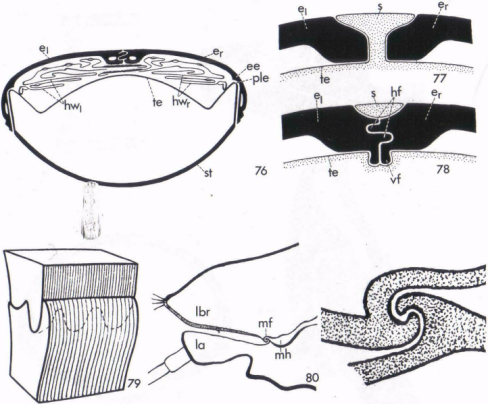
 Figure 4 – Hydroporous ferrugineus (Nachtigall p16)
76 – interlocking of abdomen and elytra
77 – interlocking of scutellum and elytra
78 – double tongue and groove joint between the elytra
-
Lamellicorn beetles have 15 clasps on their elytra.
-
Stephanolepas (a type of barnacle) has a mitre joint with deep tongue and groove connection.
-
Carabidae ground and tiger beetles are large beetles with a permanent mortis joint between elytra.
-
Cnemidotus water beetles have elytra with a locking mechanism.
-
Gygrinidae (whirligig beetles), hydrobiidae (spiral snails), haliplidae (crawling water beetles), and dryopidae (water beetles) all have elytra that are watertight.
-
Siliphidae (carrion beetle) have sealed elytra to prevent moisture loss in their arid environments.
3.1.2Releasable attachments of two matching structures
Nachtigall defines releasable attachment mechanisms as those mechanisms allowing two different structural components to be quickly coupled and decoupled. The two parts are held together firmly as long as the connection is maintained such as key-in-lock or plug-in-socket type mechanisms with an exact morphological correspondence between mating parts.
For example, the sex organs of copulating mosquitoes, dragonflies and mayflies he describes as resembling an electrical socket in functionality. They are able to maintain the connection once it has been made against the vibrations and disturbances of in-flight copulation because their sex organs have structures that match and interlock.
He groups both rigid and flexible releasable attachments together. By his classification system, the group of releasable attachments (both rigid and flexible) is made up of two main groups:
-
Connection by two complementary parts are listed below (Sub-sections 3.1.2.1 to 3.1.2.4)
-
Attachment by one specialised device. (see Section 3.1.3)
Nachtigall lists
-
plug-in-socket,
-
hook-and-eye,
-
snap fasteners and
-
multiple connections
all as mechanical analogues to some of the complementary-part devices. These are described in the next four sub-sections.
3.1.2.1Plug-in-socket
A rod-like component is introduced axially into a corresponding tube-like component. The connection is secured against 3 kinds of displacement by the use of:
-
A tongue and groove against rotation about the longitudinal axis.
-
Guide channels with matching surfaces to prevent tilting with respect to the longitudinal axis.
-
External clamping and internal anchoring to prevent displacement along the longitudinal axis. (Figure 5)
Insert your own figure.
Figure 5 – Plug and socket analogue in copulation of the midge Limnophyes pusillus (Nachtigall p29)
Some examples of organisms that have plug and socket-type joints are:
-
copulating insects such as the dragonfly,
-
crocodiles, with teeth that fit into holes in the opposing jaw,
-
the undulating ridges of large clams Tridacna,
-
the midge Tanytarsus sylvaticus, and
-
copulating yellow fever mosquito’s Aedes aegypti. (Figure 6)
.

Region of Interest Depicted Below


Stage One Stage Two Fully-docked!
Figure 6 – Egyptian mosquitoes Aedes aegypti approaching the copulatory position with the final engagement of two hook mechanisms (Evans p23).
3.1.2.2Hook-and-eye
These connections are accomplished by engaging the hook of one body part with the eye of another and they are good for attachment under tension but do not prevent tilt nor rotation.
The presence of guide grooves can prevent tilt in some species and rotation may be prevented by the presence of two hooks of the same type next to each other but with the greatest possible distance of separation between them thereby increasing the opposing rotating moment. For example, bird mites have genitalia that lock together, with a flange-capsule structure on the female dorsal surface matching capstan-like protuberances on the posterior ventral surface. In this case the female flange guides the male capstan into the correct “docking” position for insemination (see Figure 7 – Bird mite Dermanyssus (Nachtigall p35)).
 A B
A B
Figure 7 – Bird mite Dermanyssus (Nachtigall p35)
A – Bird mite Dermanyssus [IV]
B – In copulation the male slides over the top of the female to engage sex organs
female
male
C
Figure 7 cont. C – Capstan and flange of engaged bird mite sex organs (Pterophagus strictus). The capstan and flange prevents tilt but not rotation. Note that in section, the capstan has a profile that is two opposing hooks.
3.1.2.3Snap Fasteners
-
As per common engineering terminology, the male is the peg that is expanded at the end and the female is a socket of the same diameter as the peg.
-
In squid, the mantle is joined to the body by two snap fasteners. The female socket (mantle) will have an inner rim reduced slightly by some spring arrangement (such as cartilage) to maintain a seal when the muscular mantle contracts and seals to expel water under pressure down the funnel (male), for propulsion through the water. e.g. squid mantles Symplectoteuthis and Grimalditeuthis, Cranchidae, Oegopsidae. (See Figure 8 – Snap-type connection in Sepia officinalis (Nachtigall p38)
Snap connector where mantle margin meets funnel.
The g’s are two locators that locate into depressions at b.
funnel
Mantle
igure 8 – Snap-type connection in Sepia officinalis (Nachtigall p38)
-
T
Suckers modified into hooks
he tentacle suckers on the two long tentacles of some squid species have a modified system of suckers, with multiple hooks for holding prey. A system of studs and sockets on the tentacle surfaces above and below the hooked sections of each tentacle interact and connect when the prey is clasped by an opposing tentacle, thus aiding the clasping effort. See squid Onychoteuthis, Abralia, Galiteuthis.(see Figure 9 through to Figure 13)

Studs and sockets
Figure 9 – Tentacular fasteners (Nachtigall p38)


Figure 11 – Squid Onychoteuthis [VI] Figure 12 – Squid Galiteuthis glacialis [VII]

Figure 13 – Squid Galiteuthis glacialis (drawing) [VII]
3.1.2.4Multiple Connections
A mechanical example of multiple connections is a zipper, where a separate slider draws two edges of complementary morphologies together. Such sliders do not exist in nature and from the notes in Nachtigall it would seem that multiple connectors are mostly modified systems of alternate teeth, that vary greatly in modifications from simple interlocking rows of a few teeth to the interlocking of the two hemi-elytra of the north American water bug Plea striola where a great degree of interlocking is required to maintain the water tight seal when the bug is submerged.
Radiolaria are protista, unicellular organisms known for their geometric form and their symmetry because they produce a skeleton of crystal silica. They date back to the Cambrian age and some use delicate interlocking shell margins of hooks and eyes for attachment.
Shell margin
Figure 14 – Radolaria (Nachtigall p42)
3.1.3Releasable attachments by one structure
Nachtigall lists
-
clamps,
-
grippers,
-
hooks for special substrates,
-
multiple-hook devices,
-
probabilistic fasteners, and
-
expansion fastenings.
as releasable attachments by one structure, compared to the two structures of the previous section.
3.1.3.1Clamps
-
Figure 15 shows an example of split-sleeve clamps: consider the notched antenna cleaner in foreleg of honey bee. (see below)
-
Vices – e.g. pincer wasps, family Dryinidae, mantids. (see Figure 16)

Notch in tarsus for cleaning antenna by pulling thru’
Figure 15 – Antenna cleaning apparatus of honey bee (Apis mellifera) (Nachtigall p38)
1. 2.
3. 4.
Figure 16, 1-4: SEM’s of the vice mechanism of the praying mantis, zooming in on a single tooth to show surface morphology (Saunders, 2002).
-


 Forceps and medical equipment: consider the beaks of many birds (Figure 17).
Figure 17 – Clockwise from top left: 215 – an eagle, 218 – a vulture, 223 – Anarhynchus frontalis, 219 – a spoonbill (2 views) (Nachtigall p52)
They have been copied in many surgical instruments that perform similar tasks.
3.1.3.2Grippers
a) Nutcracker type
b) 4 Jaw grippers
c) Antennae of some insects have prehensile joints that can be used for gripping the mate during copulation.
3.1.3.3Hooks for Special Substrates
Hooks are used to:
-
Connect insect wings reversibly with one another
-
To attach the body of an animal to some substrate
-
To manipulate particles of food and other objects.
In the case of wing connectors (see Figure 18), Lepidopterans (butterflies and moths) have 2 types of hook mechanisms, jungate (the hooking mechanism extends from the forewing) and frenate (the hooking mechanism extends from the hindwing).
A B
Figure 18 – Wing connections jungate (A) and frenate (B) (Nachtigall p61)
f = forewing, h = hindwing, r = setae
Hooks are also used to join the forewings and hindwings of:
-
Bugs
-
Hymenopterans (bees, wasps, ants, sawflies), particularly insects with 2 pairs of wings that beat with a high frequency such as bees (Figure 19), shield moths (which both have finely matched hook mechanisms) and hawkmoths (Manduca sexta) (Figure 20 – Manduca sexta (Hawkmoth) in flight). The honeybee Apis mellifera has a single row of fine hooks (hamuli) on the costal vein of the hindwing which catch upon the undercut ridge of the posterior margin of the forewing.

Figure 19 – The connections between the wings of Apis mellifera seen from above and a honey bee in flight. (Nachtigall p61) [VIII]

Figure 20 – Manduca sexta (Hawkmoth) in flight [IX]
-
Pentatomid beetles of the genus Palomena have a snap mechanism against tensile stress worth looking at. Also Graphosoma italicum. (see Figure 21 and Figure 22)


Figure 21 – Shield bug Palomena [X] Figure 22 – Mating shield bugs Graphosoma [XI]
-
Pyrrhocoridae (red bugs, Figure 23), winged heteropteran bugs Gerridae (water striders, Figure 23 –Pale cotton stainer bug Figure 24 – Water strider Gerridae [XIII]), and Homoptera (cicadas, leafhoppers, aphids, scale insects and mealy bugs etc) similarly, all having hooking mechanisms clasping their elytra together.


Figure 23 –Pale cotton stainer bug Figure 24 – Water strider Gerridae [XIII]
Pyrrhocoridae: Dysdercus sidae [XII]
Similarly, European Ciccada Triecphora vulnerata (Cercopidae,Figure 25) and Snakeflies (Raphidioptera, Trichoptera, Mecoptera, Figure 26) and sand flies Rhyacophila dorsalis have wing connectors.


3.1.3.4Anchors
Sessile animals have special clinging or fixation organs:
The oncomiracidium (Phylum Platyhelminthes, Class Monogenea)(see Figure 27) are generally ectoparasites in the gills or body surface of fish and typically dorso-ventrally flattened, acoelomate with no anus and bilaterally symmetrical, or endoparasitic in the buccal cavity, cloaca or bladder. They have a large posterior sucker or opisthaptor as an attachment mechanism which is a disc with a double circle of hooks, each hook like a medieval halberd with a shaft, sharp cutting edge and a terminal process on one side.
-
Branchurian crustacean or carp lice have suckers and angled hooks at the base of the first pair of antenna ( Figure 28).


-
Blood-sucking isopods: Gnathia live on fish and have boat-hooks and harpoon hooks to grasp host fish and attach themselves to them (Figure 29).

Figure 29 – Gnathia maxilaris [XVIII]
-
Kalyptorhynchia (turbellarian worms) have conical pincers which have a pair of hooks at the tip which they dig into the flesh of prey.
-
There are also instances of two rows of hooks acting in opposition to each other, for example, Sea stars (Pectinate pedicellariae).
-
Peacock, heron, dipper and woodpecker tongues are horny with edges with keratinised teeth. These teeth are saw-like and serve to prevent the prey working loose when they are speared by the tongue. (Figure 30)

Figure 30 – Woodpecker tongue showing keratinised teeth [XIX]
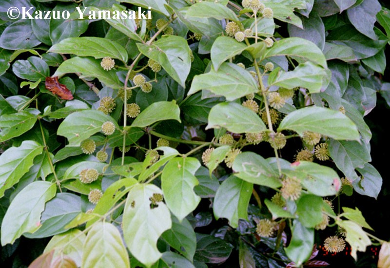
There are many climbing plants of which the most successful make use of spines or hooks to attach themselves to the host plants.
-
Gleichenia linearis is a tropical rain forest fern and palms of Plectocomia (Figure 31).

Figure 31 – Plectocomia himilayana showing climbing spines [XX]
-
Uncaria have climbing hooks on their stems that anchor into rough or mouldering substrates (Figure 32).

Figure 32 – Uncaria with the red arrow indicating typical hook position [XXI]
-
The stalk of the hop plant has longitudinal climbing hooks and the runner bean has climbing hairs in the shape of crampons.
3.1.3.5Multiple hook Devices
-
Trypanorhyncha (tapeworms of the cestode order) (Figure 33) have arrays of hooks, macro-hooks, micro-hooks, micro-hooklets and hook chains.

Figure 33 – Tapeworm trypanorhyncha lascisthynchus [XXII]
3.1.3.6Probabilistic Fasteners (random hooking or “burr”- type devices)
Nachtigall has divided this group into devices obeying
-
The Burr Principle (having hooks),
-
The Comb Principle (no hooks),
-
The Feather Principle (barbs and barbules) or
-
The Micro-hook Principle (fields of tiny hooks)
These devices are described overleaf:
-
-
Burr principle
-
-
Species Arctium lappa (burdock) ( Figure 34)
-
Agrimonia eupatoria The fruits of both of these consist of round fruits with rings of barbs having long shafts and single barbs. ( Figure 35)


-
Cynoglossum spine tips have a double anchor.
-
Galium (bedstraw) has thousands of fine barbs on its climbing stem.(Figure 36)

Figure 36 – Bedstraw Galium [XXV]
-
Bellostoma stouti (a type of fish) lays eggs that have bundles of hornlike threads at each end with “buttons” at the tip. In water these anchor filaments extend to catch the hooks of nearby eggs.
-
Acanthocephalan worms have a proboscis that can invert so that the ring of hooks points inwards. When internal pressure is increased the teeth appear “at the margin” and are turned to the outside where they catch into the villi of the host’s gut. It uses a peristaltic action to manoeuvre through the gut. (Figure 37)

Figure 37 – Echinorhynchus salmoides acanthocephalan worm [XXVI]
-
Kinorhyncha (of the class Aschelminthes) have a crest of hooks on their heads. They use this as a “mud” anchor which anchors them to or helps them to move about, on the sea floor. See also echiuroid worms.
-
Cat and cow tongues as a brush and curry-comb since the tongue is covered in fields of “re-curved, horny teeth”.
-
-
The Comb Principle
-
Consider rough hair being combed with a very fine tooth comb. The comb snags because hairs running at angles to one another become entwined. This principle is used in ectoparasitic insects living in the fur of vertebrates. The combs are called ctenidia and are one or more rows of closely and evenly spaced bristles. For example:
-
Siphonaptera (fleas)
-
Nycteribiidae (bat flies)
-
Polyctenidae (bat bugs)
-
Platypsyllus castoris (beaver beetle)
-
-
Feather Principle
-
The vane of a feather consists of a number of side branches called barbs. Each barb has “second order side branches” called barbules.
A feather vane can be broken up into segments by pulling in a direction perpendicular to the barb longitudinal axis.
Figure 38 – a) Hook barbule b) Bow barbule c) Probabilistic fasteners on hook and bow barbules interlocked (Nachtigall p75)
One side of the barbule has hook barbules and the other has rows of bow barbules. It is a probabilistic fastening with no particular matching of elements required. The vane is allowed to separate under strong local pressures into segments because otherwise the vane would be destroyed. (Figure 38)
The hook barbule has small “inward curving spurs at its distal end” to prevent the attachment from separating by axial sliding.
-
-
Micro hook Principal
-
Geckonidae and some Iguanidae (Anolis) have “leaf-like broadenings of fingers and toes called digital pads or discs” (p78) (even on the tip of the tail sometimes). The surfaces of these pads have transverse lamellae that are covered in dense rows or brushes of tiny bristles. Increased blood pressure in the capillary system of the pads causes the brushes to be pressed firmly against the substrate. Each bristle in turn has a tuft of hundreds of minute processes with tips bent like hooks. These hooked tips are thought to interact with the most microscopic of surface irregularities and to utilise forces of an electrical (surface atomic and molecular charges) or capillary (gluing with water) nature.
4Classification by Function – Gorb
Gorb follows a classification definition based on functionality. For the class of insect cuticle attachment devices he defines the functions:
-
Hooking to the substratum
-
Animal associations: phoresy (the behaviour of animal dispersal using other animals), parasitism, predation
-
Hooking within biological tissues
-
Attachment during copulation
-
Interlocking of body parts
Figure 39 below illustrates 8 fundamental classes of fixation principles including hooks, lock or snap, clamp, spacer, sucker, expansion anchor, adhesive secretions, friction.

Figure 39 – Eight fundamental classes of fixation principles: hooks (A), lock or snap (B), clamp (C), spacer (D), sucker (E), expansion anchor (F), adhesive secretions (G), friction (H) (from Gorb p38)
Gorb concludes his description of the functions of hooks in insect species with the following description (p50):
“The hook mechanism is usually comprised of two complementary surfaces. These surfaces are not necessarily mirrored copies of each other but some dependence on the corresponding surface does exist. If both surfaces bear hooks (wing-interlock) their dimensions are usually predefined in order to optimise attachment and the probability of attachment as well. When only one surface bears real hooks, they could only attach efficiently to a particular range of textures (tarsal claws, hooks of phoretic and parasitic animals). The hook design can range from unicellular acanthi and multicellular setae to spines and cuticular folds.”
4.1.1Hooking to the substratum
This function is viewed in terms of claws to aid locomotion or to provide anchorage. For example in terrestrial locomotion the tarsal claw/s interlock with the surface textures to generate friction. Orb-web spiders have claws with a comb-like serrate bristle edge. This plays an important part in interlocking on the silk thread. Some butterfly pupae (e.g. Thyridopteryx ephemeraeformis, Lepidoptera, Psychidae) have hooks at their posterior end to anchor them inside their cocoon. Hooks are also used to attach material to an animal body (the crab Loxorhynchus crispatus decorates its shell by hooking with specialised setae).
4.1.2Animal associations: phoresy, parasitism, predation
Bird mites (Order Acari genus Michaelicus) parasitize onto bird feathers. They have an asymmetric design of their legs with only one leg having a tarsus with a claw, which they feed through the barbules of the feathers to find anchorage. Hook-like devices are found only on mites parasitizing onto the stiff parts of the feather whereas mites that live on the soft parts of the feather use a clamping device.
Copepoda parasitica are parasitic copepod crustaceans with hooked appendages for attachment to host such as some species of fish lice that use hook-like appendages to attach themselves to their hosts.
4.1.3Hooking into biological tissues
Gorb lists copepod crustaceans, ixodid ticks and dipteran insects as having mouthparts with hook-like structures for attachment to hosts. Also parasitic copepods such as Hatschekia pseudohippoglossi and Trebius clidodermi.
4.1.4Attachment during copulation
Gorb adds the attachment structures of Harpocera thoracica to the organisms mentioned previously in Section 3.1.2.2.
4.1.5Interlocking of body parts
This category mainly includes wing-connectors. (Figure 40 )

Figure 40 – Wing inter-lock devices in Heteroptera and Auchenorrhyncha (Gorb p45)
5Conclusion
The preceding summaries are somewhat perfunctory. This is because they do not have much more than an illustrative and recording function. Should the opportunity arise for further research this should suffice for a first reference point. Clearly more figures could be added but time constraints mean that this will have to be left to a later date.
6References
-
“Biological Mechanisms of Attachment, The Comparative Morphology and Bioengineering of Organs for Linkage, Suction and Adhesion”, W Nachtigall, 1974 translated by M A Biederman-Thorson, Springer-Verlag, ISBN 3-540-06550-4
-
“Attachment Devices of Insect Cuticle”, S Gorb, 2001. Kluwer Academic Publishers. ISBN 0-7923-7153-4
-
“Insect Biology A Textbook of Entomology”, H E Evans, 1984, Addison-Wesley, ISBN 0-201-11981-1
-
“Plant Form An Illustrated guide to Flowering Plant Morphology”, A E Bell, 1991, Oxford University Press, ISBN 0-19-854219-4
7Image References
-
http://www.aquaculturemag.com/siteenglish/printed/archives/issues03/03articles/HeckmanFeatureForWeb.pdf (figure 1)
-
http://www.enature.com/fieldguide/showSpecies_LI.asp?imageID=19348 (figure 4)
-
http://medent.usyd.edu.au/fact/birdmite.html (figure 7)
-
http://tolweb.org/tree?group=Abralia&contgroup=Enoploteuthidae (figure 10)
-
http://tolweb.org/tree?group=Onychoteuthis&contgroup=Onychoteuthidae (figure 11)
-
http://tolweb.org/tree?group=Galiteuthis&contgroup=Cranchiidae (figure 12&13)
-
http://www.floridanature.org/species.asp?species=Apis_mellifera (figure 19)
-
http://zebra.biol.sc.edu/moth/manduca.html (figure 20)
-
http://www.bioimages.org.uk/HTML/T1457.HTM (figure 21)
-
http://www.geocities.com/pelionature/Graphosoma_italicum2.htm (figure 22)
-
http://insects.tamu.edu/fieldguide/aimg60.html (figure 23)
-
http://www.insectimages.org/browse/family.cfm?id=Gerridae (figure 24)
-
http://eny3005.ifas.ufl.edu/lab1/Homoptera/Homoptera.htm (figure 25)
-
http://www.lib.ncsu.edu/agnic/sys_entomology/taxon/raphidiodea/#order (figure 26)
-
http://bioweb.uwlax.edu/zoolab/Table_of_Contents/Lab-4a/Class_Monogenea/class_monogenea.htm (figure 27)
-
http://www-biol.paisley.ac.uk/courses/Tatner/biomedia/pictures/fishl.htm (figure 28)
-
http://www.konig- photo.com/english/galerie/zoom.asp?pre=8206&NumPhoto=8207&suiv=8208&Rub=483 (figure 29)
-
http://www.hiltonpond.org/ThisWeek030308.html (figure 30)
-
http://www.palmsoftheworld.com/plec.htm (figure 31)
-
http://www.pharmakobotanik.de/systematik/7_bilder/yamasaki/Uncaria.jpg (figure 32)
-
http://www.mic-d.com/gallery/darkfield/tapewormscolex.html (figure 33)
-
http://botanical.com/botanical/mgmh/b/burdoc87.html (figure 34)
-
http://www.bioimages.org.uk/HTML/P146161.HTM (figure 35)
-
http://plants.usda.gov/cgi_bin/large_image_rpt.cgi?imageID=gaap2_002_avp.tif (figure 37)
-
http://www.inhs.uiuc.edu/chf/pub/surveyreports/nov-dec96/acanth.html (figure 38)
